
Welcome to the research testing information page. On this page you'll find information for utilizing the pathology laboratories as part of your research project.
Any researcher utilizing the Department of Pathology's services for a study needs to obtain prior approval from the department. The approval process starts by the researcher including these items:
A completed Research Application for Utilizing Pathology Services (RA Path) form
- The protocol or abstract and if available, pertinent laboratory manual
- The IRB application
- The consent form used to obtain participate approval
- The face page of the IRB approval
All five are required for approval but to prevent delay, you may initially submit (1) through (4) above to Pathology Research Coordinator. If you submit using the Submit button, a window will open where you can attach your protocol or other documents, enter identifying information in the subject line, etc.
Additional information and forms:
- Common Pathology Services Price List
- Application for Digital Scanning
- Application for DeGowin Blood Center Products
- Articles about Leukocyte Reducation System (LRS) Filters
Viewing the PDF forms in web browsers may limit functionality, please download each form to complete.
House staff research support
The opportunity for residents and fellows to perform research under the direction of a faculty mentor is an important part of house staff training.
Since research projects carried out by a trainee-faculty team are often conceived at the initiation of a rotation or fellowship, they can be difficult to plan for during the annual budget cycle. Applications can therefore be submitted throughout the year.
Please be aware that the Pathology Laboratories are primarily for patient care. Patient specimens will take priority over research specimens.
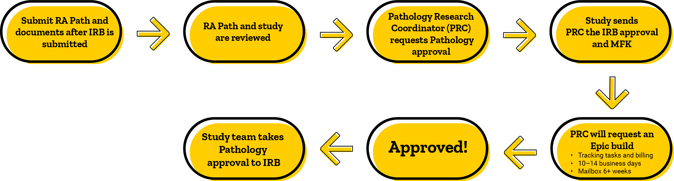
Approvals timeline
Please note this is an estimate, please plan accordingly. It is highly recommended to apply for pathology approval when the IRB application is submitted.
Billing information
If you choose reporting Option 1 on the RA Path form: The research results will be displayed in Epic under the participant’s UI Health Care medical record number (MRN) in his/her electronic health record. To ensure correct billing of this research testing, the principal investigator must contact Research Billing Compliance in Patient Financial Services (PFS).
If you choose Option 2 or 3 on the RA Path form: Invoices are generated through Pathology’s UI Diagnostic Laboratories (UIDL) billing system with monthly billing to your research MFK. Test ordering is not done via standard Epic processes; the laboratory uses a special research ordering process. You will receive research results in 1 of 2 ways:
- Automated faxing to your designated, secure fax number
or - e-Fax associated with an Outlook shared mailbox
Translational research core labs and services
Histology Research Laboratory (HRL) and Comparative Pathology Laboratory (CPL)
The Histology Research Laboratory (HRL) and Comparative Pathology Laboratory (CPL) provide necropsy, gross sectioning of research tissues. They are fully equipped to provide tissue fixation and cryopreservation as well as histological needs like tissue processing, embedding, and staining. HRL works with studies to provide special stains and IHC stains. HRL/CPL has CLIA accreditation.
CPL contact information:
HRL contact information:
Tissue Procurement Core (TPC)
As a CAP accredited biorepository, the Tissue Procurement Core (TPC) collects and stores human tissues and specimens. We encourage the use of TPC to assist studies in collecting various specimens because their workflows are embedded within clinical pathology’s workflows. TPC can collect and store fresh tissue, blood, urine, and DNA. They can assist in providing consented specimens for various projects through the BioMER.
TPC contact information:
Contact information
When using UI Health Care Pathology Laboratories to perform services and testing specified by a sponsor's research protocol, you may be asked to provide the sponsor with documentation of the laboratory's licensure, certifications, and accreditations.
If you would like more information or have questions, please contact Pathology Research Coordinator.

Jessica Cross, MPH
Definitions/terms/vocabulary words:
Research pathology
- PRC: Pathology Research Coordinator (pathresearchcoord@healthcare.uiowa.edu)
- Client ID/submitter: a number assigned to the study during intake to identify the study.
- Bylaw wavier: is needed when fresh tissue is being requested, a letter from the study to the pathology department requesting fresh tissue, pathology issues a bylaw waiver to the study team confirming what is allowed to be collected
- Letterhead: printed heading on stationary stating a person’s or organization’s name, address, and phone number. Placed in the top right corner of a letter.
- Archival tissue: tissue that has been processed and embedded in an FFPE block
- Fresh tissue/wet tissue: this is tissue that has not been preserved yet.
- Fresh tissue needs to be handled quickly to preserve the integrity of the tissue structure. Since there is no blood flow to the tissue, degradation happens quickly which does impact testing and results.
- Saline is used to keep tissue moisturized in transit.
- Formalin fixed: formalin is a derivative of formaldehyde; this is the most commonly used fixative for clinical tissue.
- RA Path: Research Application for Utilizing Pathology Services
- This is used to request pathology approval
- Please provide information and materials requested on this form
- Download RA Path form
- DBC: DeGowin Blood Center
- Duration of study: the time frame you will be requesting pathology services
- MCU: Medical Center University
- MCNL: Medical Center North Liberty
- MCD: Medical Center Downtown
- IRL: Iowa River Landing
Surgical pathology
Surgical pathology: tissue from a biopsy or surgical resection is removed from the surgical/procedure field and sent to Surgical pathology for gross description and dissection.
Gross description and dissection: includes a description of the tissue received (color, texture), measurements, inking margins (if applicable), submitting all tissue or representative tissue in cassettes for histology processing.
Sectioning: pathologist assistants will dissect the specimen, making 3mm cuts (bread-loafing) to fit the tissue in a cassette(s).
Cassette: a small rectangular plastic container with perforations to allow processing chemicals to infiltrate the tissue inside.
Surgical number: this is a case number that is assigned to the patient’s specimen by the surgical pathology team for identification.
- S# - surgical pathology
- NG# - cytology
- H# - bone marrow
- A# - autopsy
Punch biopsy: a cylindrical tool is used to press into the affected area (like a cookie cutter) and remove a small piece of tissue; normally this is done on the skin.
Resection: a surgical procedure removing a large portion of affected tissue or the entire organ. An example would be a lung wedge, breast lumpectomy, or removing a section of colon.
- This is normally done as part of treatment to the patient’s diagnosis.
Pathology report (path report): this includes the gross description from the pathologist assistant, what areas of the specimen were sampled, how much tissue was submitted, the pathologist’s interpretation, add-on testing, special stains, cancer staging.
Biopsy: small piece of tissue from the affected area taken to diagnose/confirm diagnosis.
- FNA biopsy: fine needle aspirate biopsy, a narrow diameter gauge (fine gauge) needle is used to sample the affected area. Small cylindrical fragments of tissue are removed from the patient and sent to surgical pathology for processing.
- Core biopsy: a larger gauge needle is used to sample the affected area.
- Excisional biopsy: this is a larger chunk of tissue that is removed from the body.
- Examples of this would be an ellipse of skin removed to diagnose and or treat skin cancer.
- Shave biopsy: taking a small layer of tissue off the affected area; normally this is done on the skin surface to remove a mole or suspicious lesion.
Histology
Histology processing of tissue: Histology techs will place the cassettes containing tissue on a processing machine that treats the tissue with various chemicals for preservation and to enhance stain uptake.
Embed/embedded/embedding: to be able to cut tissue on to a slide it needs to be held within a medium to help keep the tissue structures and integrity stable, we do this by embedding tissue in paraffin wax. Much like olive loaf (olives held in suspension by “meat”).
FFPE block: formalin fixed paraffin embedded tissue block.
- Clinical block: FFPE block created from the patient’s clinical care biopsy or surgical resection
Microtome: cutting tool/machine that histology techs use to slices the FFPE block very thin (standard is 4-5µm), think of a meat slicer at the deli cutting the olive loaf really thin.
- thickness: refers to the depth of the cut from the FFPE block.
- Scrolls/curls: are a thicker cut from the FFPE block, i.e. 10µm. These are thick enough cuts that the tissue rolls over on itself like a pill bug rolling up.
Slides/histology slides: piece of elongated glass used to adhere the FFPE tissue cut by the microtome.
- Histology will embed tissue into paraffin wax, once the wax is set they will take the block to a microtome, place the block in this machine and cut slides from it. The cut piece is floated in a water bath, the slide is submerged on an angel in the water bath, moved under the tissue cut and as the histology tech lifts the slide, the piece of tissue will adhere to the slide.
- Plus slide/positive charged slides/charged slide: chemically treated slide to promote tissue adhesion to the slide.
Unstained slide: routinely this is a plus slide with tissue adhered to it that is requested by research studies. The study team sends the slides to the central lab and they will stain the slides with their desired stains and techniques.
H&E stain: hematoxylin and eosin stain, this is the most routine stain done in pathology.
- Other stains and staining methods highlight different structures of the tissue.
TMA: tumor microarray
- An H&E slide is made from the requested block. The pathologist will circle the area on the H&E slide that needs to be extracted from the block. The histology tech will look at the marked slide and line that up with the block. They will insert the punch tool to extract the area that was marked. The core from that punch will be embedded in a new block for research. This normally is done using multiple blocks to create an array of target areas from different blocks represented in one block. This TMA block is then cut using the microtome to make a slide. The slide now has small circular representative areas from multiple cases that are stained. Now the pathologist has one slide they can review multiple cases of the same disease type.
- Punch: same technique as a punch biopsy though this is a punch to an FFPE block. The punch tool has various diameters to accommodate the affected area to be sampled. The devise is pressed into the marked area of the FFPE block resulting in a punch. This punch is then embedded into a block
- Multiple punches embedded into a block is a TMA
- HRL can accommodate an 8 x 10 TMA
Exhausting a block: taking all the available tissue from the FFPE block to make slides.
- Based on the tissue sample size, we can only get so many slides made from the available tissue. Here as well as at other facilities, hospitals, pathology departments, have a responsibility to the patient to manger the tissue that was retrieved during a biopsy or surgical procedure. We do everything we can to preserve the FFPE block and take only what we need to assist in the patient’s diagnosis and care. Through approval from pathology, research studies are allowed to take more tissue from the block to create slides for their projects however, they cannot exhaust the clinical block.