University of Iowa Health Care’s rare “brain bank” has contributed to a new study published in Nature that shows that repetitive head impact (RHI)-related brain injuries result in brain cell loss, inflammation, and vascular damage in young, former contact sport athletes. Importantly, many of these detrimental changes were seen in young athletes before the onset of chronic traumatic encephalopathy (CTE), a degenerative brain condition that leads to problems with thinking and emotional regulation. The study was led by researchers from Boston University (BU) Chobanian & Avedisian School of Medicine.
Repetitive head injuries, like those sustained in contact sports, are a key risk factor for developing CTE. However, the biological and cellular pathways damaged by RHIs and how that damage might lead to the neurological and cognitive problems of CTE are not clear.
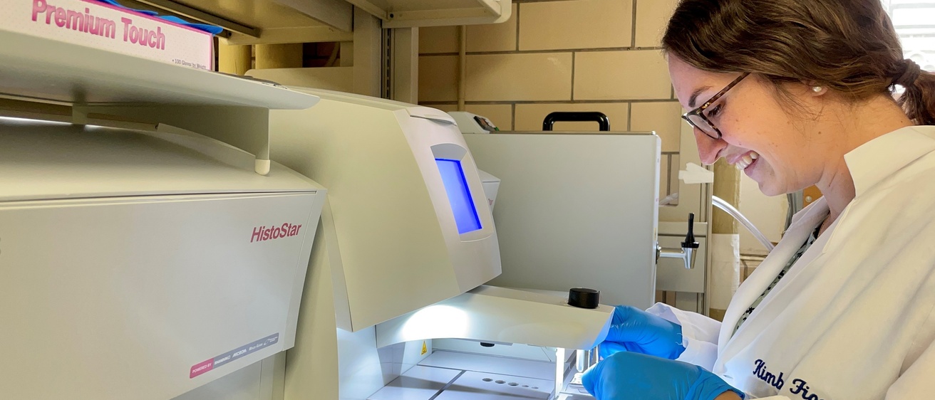
The Iowa Neuropathology Resource Laboratory is a central hub for resources related to neuropathology research and education and serves as the brain bank for the University of Iowa.
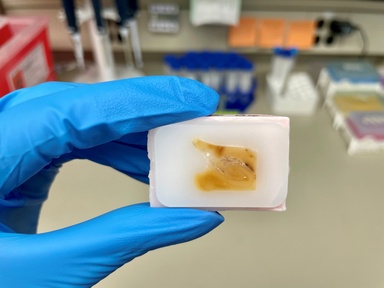
The UI brain bank, or Iowa Neuropathology Resource Laboratory (INRL), is led by UI pathology researchers Marco Hefti, MD, and Kimberly Fiock, PhD. It is a rare resource — there are fewer than 200 brain banks worldwide — which catalogs, stores, and distributes donated brain tissue to researchers studying how our brains work and are affected by disease. The INRL provided the BU study team with rare brain samples from younger individuals who did not have RHI damage. These samples served as critical age-matched controls, allowing the researchers to investigate the cellular and molecular changes in brain cells initiated by RHI.
To identify the earliest changes from RHI, researchers performed single nucleus RNA sequencing on samples of frozen human brain tissue from 28 men between the ages of 25 and 51. The samples were divided into three groups: the INRL control samples from eight men who didn’t play contact sports and did not have a history of head injury; brain tissue from an RHI group comprised of eight American football players and a soccer player, none of whom were diagnosed with CTE; and a CTE group of 11 contact-sport athletes with low-stage (defined as Stage 1 or 2) CTE. All results were further validated and confirmed in larger sample sets and through comparison to other published studies.
As expected from previous research, the brains of athletes diagnosed with low-stage CTE had significant inflammatory and vascular changes. However, this study also found similar levels of vascular injury and inflammation in athletes without CTE, suggesting that RHI-related brain injury is not solely dependent on CTE.
Strikingly, there was a 56% loss of neurons in young athletes participating in contact sports. The neurons were lost from the brain regions, known as cortical sulcal depths, that undergo the highest mechanical forces during head impact injury, and where CTE first develops. Neuron loss was observed in all athletes, regardless of whether they had CTE.
“This study looked at several different subtypes of cells involved in neurodegenerative disease processes and severe traumatic brain injury and, for the first time, reported these same changes in a younger cohort exposed to repetitive head injury,” says Fiock, director of the INRL. “The findings suggest that exposure to repetitive head injury on its own is enough to cause significant neuronal loss and dysfunction, even in the absence of CTE pathology. This means that the risk of playing contact sports is not just about the development of CTE, but rather, the fact that it can cause significant, long-term damage to the brain from the impact itself.”
Editor’s Note: this article is adapted from a Boston University news release